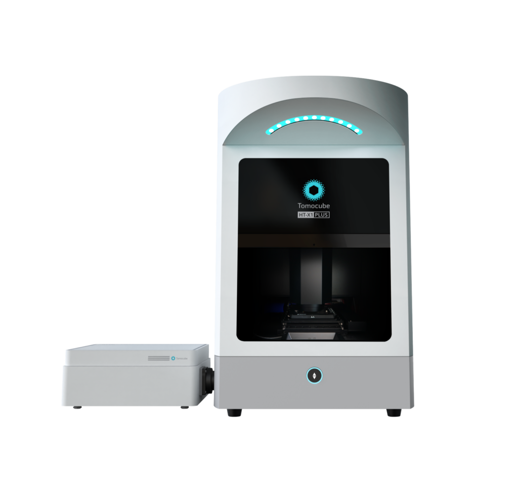
Tomocube Introduces the NEW HT-X1 PLUS Holotomographic System

Setting New Standards in 3D Imaging Throughput for Biomedical Screening

We’re thrilled to announce the official launch of the new HT-X1 Plus system, the latest innovation in the high-performance holotomography portfolio from Tomocube. Continuing to push the boundaries of bio-imaging, this new model takes everything you loved about the HT-X1 to the next level. Whether you’re advancing biomedical research or tackling new imaging challenges, the HT-X1 Plus opens up a world of possibilities.

Why you’ll love it!
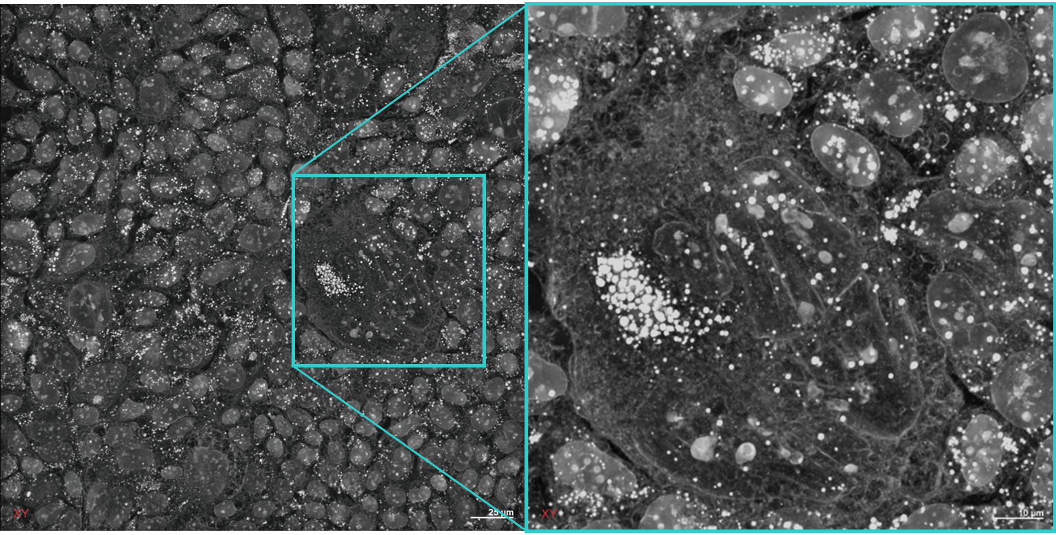
High content screening of live cells and organoids

The HT-X1™ Plus is optimized for high-throughput screening, making it highly suitable for high-content, image-based drug screening research.  
https://stratus.campaign-image.com/images/153213000054294155_zc_v1_1727123765449_629de15c_0503_8b6c_52ea_f97154bf6ed7.jpeg
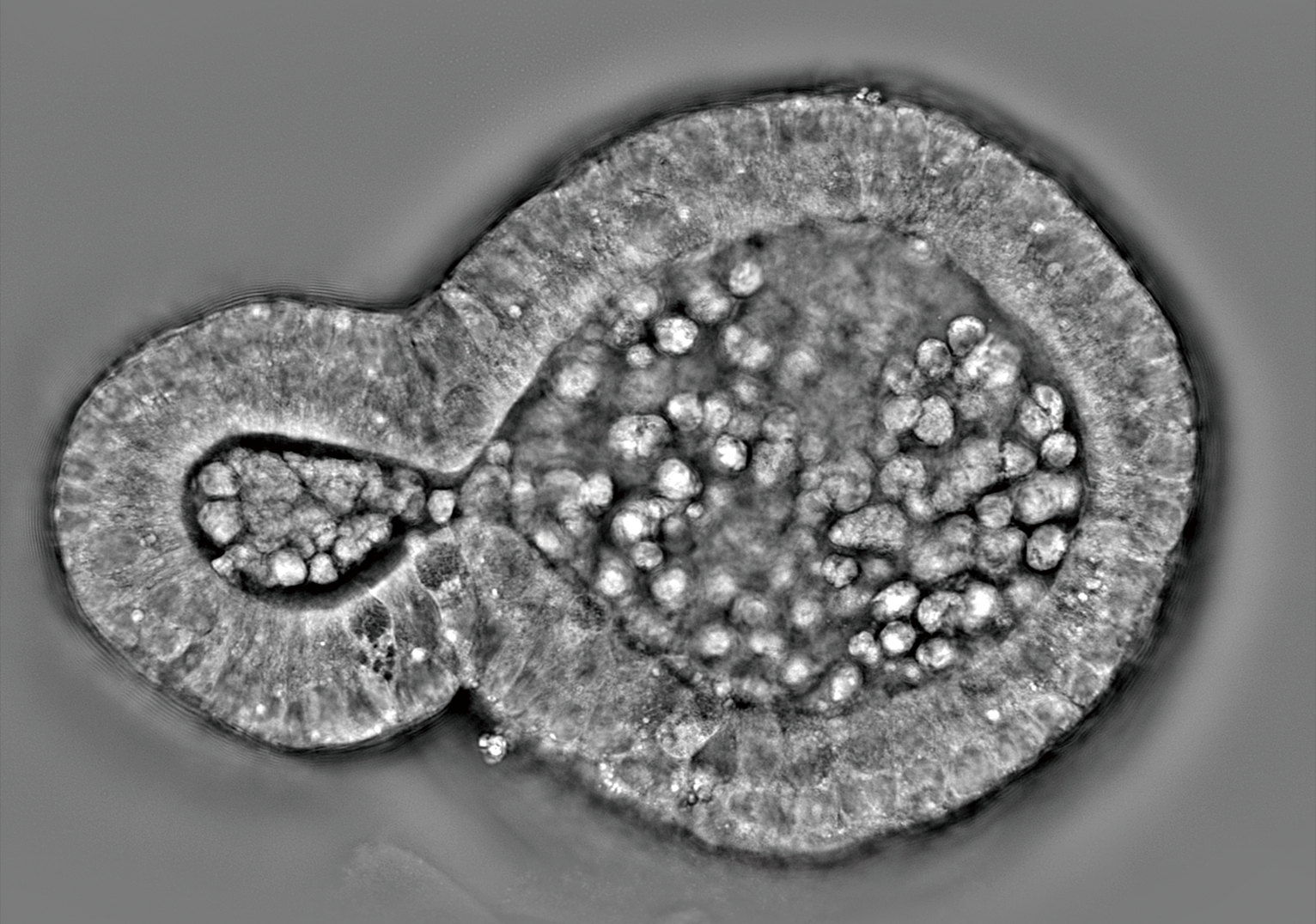
High resolution imaging of 3D biological samples 

The new features of the HT-X1™ Plus are especially advantageous for research involving 3D cultures, enabling the detailed investigation of dense organoids and intact tissue sections.
https://stratus.campaign-image.com/images/153213000054294155_zc_v1_1727123765449_629de15c_0503_8b6c_52ea_f97154bf6ed7.jpeg
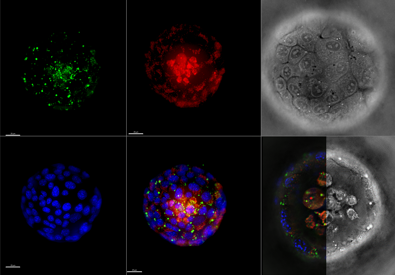
Enhanced Correlative Fluorescence Imaging

The HT-X1™ Plus offers enhanced multimodal imaging capability with its fluorescence module (FLX™) featuring an sCMOS camera designed specifically for precise signal intensity measurements.   
https://stratus.campaign-image.com/images/153213000054294155_zc_v1_1727123765449_629de15c_0503_8b6c_52ea_f97154bf6ed7.jpeg
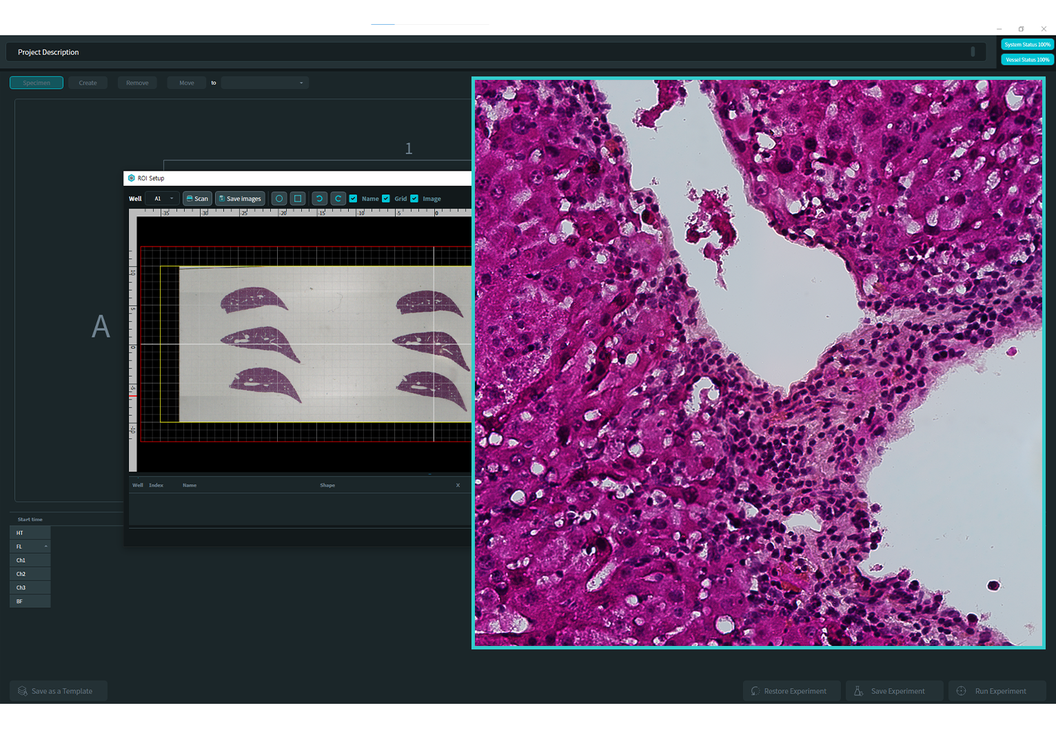
Color Bright-Field Imaging for Histological Studies

With the new color bright-field imaging modality and wide preview scan features, researchers can gain deeper insights into tissue section studies. 
https://stratus.campaign-image.com/images/153213000054294155_zc_v1_1727123765449_629de15c_0503_8b6c_52ea_f97154bf6ed7.jpeg
Interested in this new product? Let us know >>

PHENOM SEM on TOUR 2024

MAY – NOVEMBER 2024

6 Countries, 18 locations

Our one-day free seminar offers innovative solutions and practical experience for electron microscopy in your area!

The largest and for years successful “Phenom REM on Tour” event in Europe will tour through 6 different countries and 18 different cities from May to November to introduce you to the innovative technology of desktop electron microscopy.

These events are of course non-binding and free of charge.

Click here for further Information and Registration:

Deutschland  Österreich  Schweiz (DE)  Schweiz (FR)  Belgien  Niederlande  Frankreich


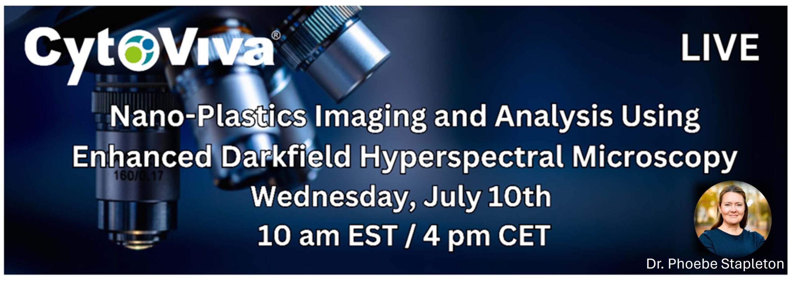
📢 CytoViva, Inc Webinar on Nano-Plastics Imaging and Analysis Using Enhanced Darkfield Hyperspectral Microscopy

You are invited to register for CytoViva´s free webinar, featuring Dr. Phoebe Stapleton of Rutgers University.

📅 Wednesday, July 10th, 10 am EST / 4 pm CET

𝗥𝗲𝗴𝗶𝘀𝘁𝗿𝗮𝘁𝗶𝗼𝗻 𝗟𝗶𝗻𝗸 👈

𝗪𝗵𝗮𝘁 𝘆𝗼𝘂 𝘄𝗶𝗹𝗹 𝗹𝗲𝗮𝗿𝗻:
The study of plastics and their impact on humans and environment is a major focus of research funding agencies worldwide. To support these studies, Enhanced Darkfield Hyperspectral Microscopy has emerged as a critical tool for label free imaging and spectral characterization of nanoscale plastics in a wide range of matrices. In this webinar you will learn the principles behind Enhanced Darkfield Hyperspectral Microscopy. This will include details on its ability to provide label free imaging and spectral characterization of nano and micron scale plastics in a wide range of in-vitro and ex-vivo environments.

Additionally, Dr. Phoebe Stapleton, Associate Professor at the Rutgers University Ernest Mario School of Pharmacy (EMSOP) will present data that focuses on identifying how nano and micron scale plastic particle exposure during pregnancy may affect the expectant mother, developing fetus, and the health of the next generation. Dr. Stapleton’s lab has utilized CytoViva, Inc’s technology extensively in this work and will show examples of these research efforts.

Join us to learn how you can use darkfield hyperspectral microscopy to advance your work researching nanoscale plastics in a wide range of complex biological environments. 👌


Schaefer-Tec Application Days 2024

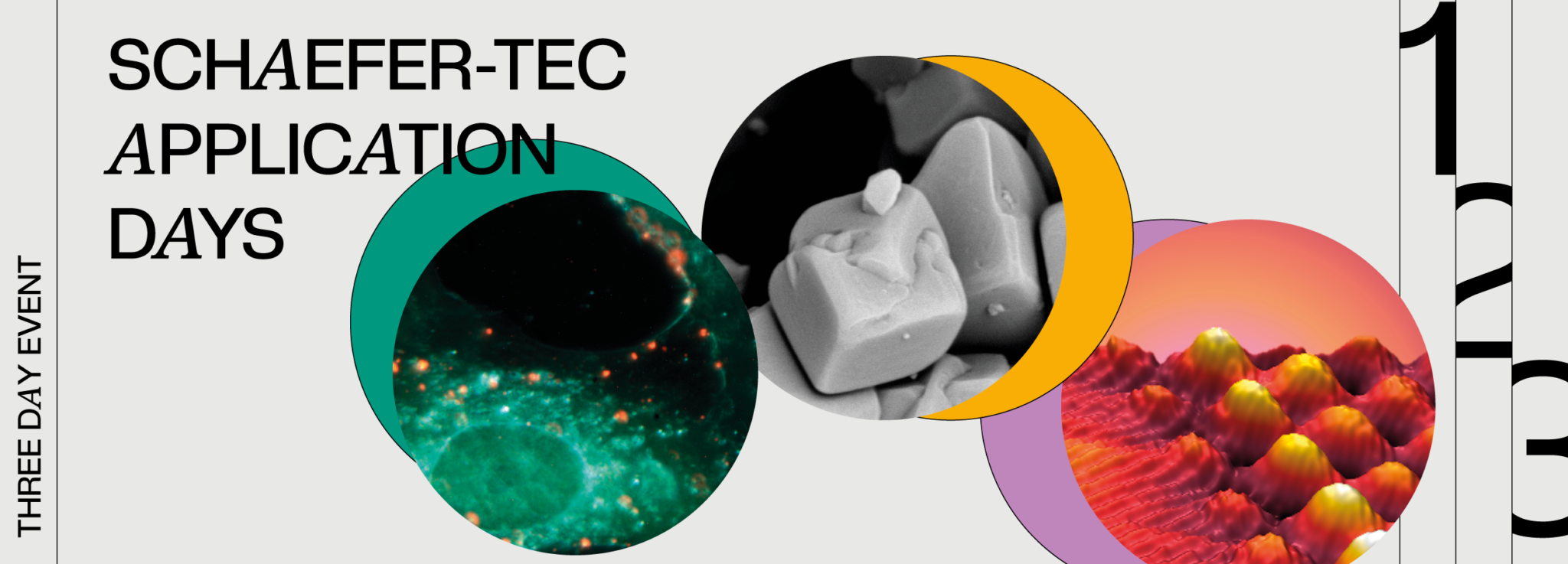
We are delighted to invite you to attend our upcoming Schaefer-Tec Application Days event, which will take place in May 2024 at our headquarters in Langen near Frankfurt am Main, Germany.

In three consecutive days, we will present the core of our product portfolio consisting of different microscopic and imaging technologies in the field of Material and Life Sciences. 

Company representatives together with invited scientists from various continents will introduce their solutions and experiences via short presentations and on-site demonstrations. This is a great opportunity to explore cutting-edge scientific instruments in action and find the right tool for your applied research and your department requirements. 

This event is free of charge, lunch and coffee breaks are included for all participants.


PRACTICAL DETAILS

Date & Time: 14th – 16th May 2024 , 8 am to 5 pm

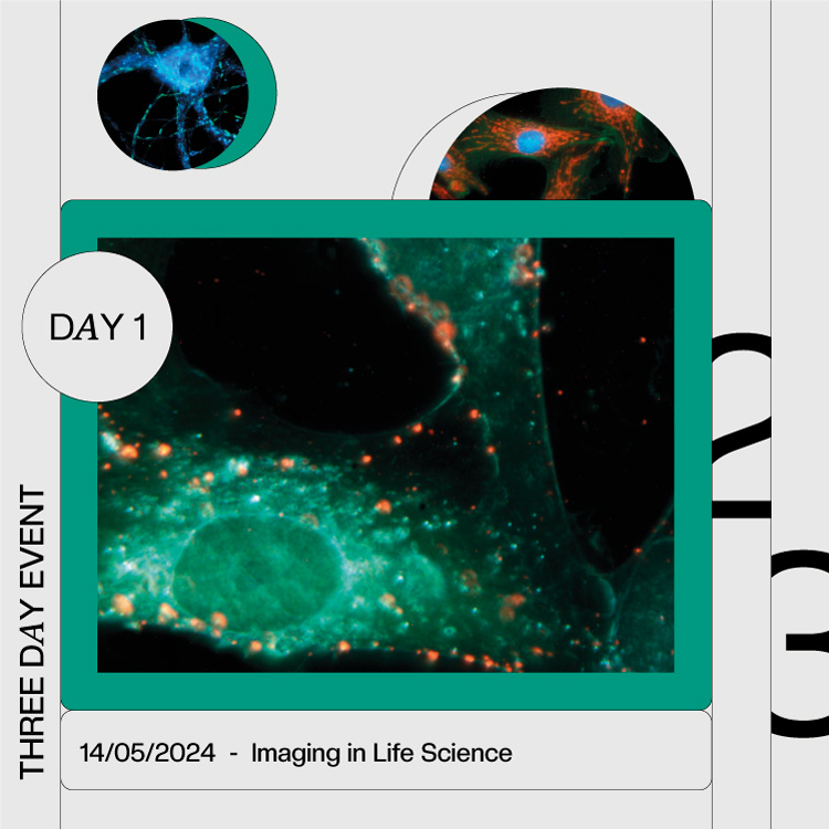
  • May 14: Imaging in Life Science
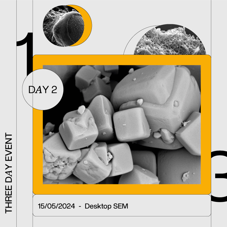
  • May 15: Electron Microscopy
  • May 16: UHV- Scanning Probe Microscopy

>>  Location: Robert-Bosch-Straße 31, 63225, Langen, Germany


WHY ATTEND THIS EVENT? 

Discover firsthand the expertise of company representatives and invited scientists from around the world as they share their solutions and experiences through short presentations and on-site demonstrations. This event offers a unique opportunity to witness cutting-edge scientific instruments in action and to find the perfect tool for your applied research and departmental requirements. 

ON-SITE SAMPLE ANALYSIS 

We offer the exclusive option for our attendees to bring samples (applicable for the 1st and 2nd day) for real-time analysis on the instruments available.  


SUPPLIERS REPRESENTED AT THIS EVENT


3 DAYS PROGRAM

DAY 1: IMAGING in LIFE SCIENCE

PROGRAM DAY 1

9:00 – 9:15Welcome & Agenda
9:15 – 9:45CytoViva Enhanced Darkfield-Hyperspectral Microscopy & Imaging Solutions
9:45 – 10:15Tomocube Holotomographic Microscopy & Imaging Solutions
10:15 – 10:45TissueGnostics Contextual Tissue Cytometry & Imaging Solutions
10:45 – 11:15Coffee Break
11:15 – 11: 30CytoViva Technology Applications
11:30 – 11:45Tomocube Technology Applications
11:45 – 12:00TissueGnostics Technology Applications
12:00 – 13:00Lunch Break
13:00 – 17:00Hands-On Demonstrations, Coffee Break & Networking 
From 17:00 Farewell-Ceremony for the Retirement of Dr. Gertrud Goetz (Managing Director, Schaefer-Tec)

HOST DAY 1

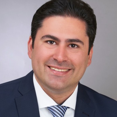
Dr. Shahriar Dantism
Technical Sales, Nanobiotechnology


DAY 2:  ELECTRON MICROSCOPY / DESKTOP SEM

PROGRAM DAY 2

9:00 – 9:15Welcome & Agenda
9:15 – 9:30Introduction of Participants
9:30 – 9:50Introduction to SEM (Lars Jansen) 
9:50 – 10:10Introduction to Luxor Sputter Coater (Jan De Munck) 
10:10 – 10:30SEM Applications (Lars Jansen) 
10:30 – 11:00Coffee Break
11:00 – 11:20Sample Preparation with Ion-Milling (Martin Schaefer)
11:20 – 11:40ParticleX – Technical Cleanliness (Dr. Sebastian Wex, RJL) 
11:40 – 12:00Nanoprobing (in SEM) with Imina miBots (Martin Schaefer) 
12:00 – 13:00Lunch Break
13:00 – 17:00Hands-On Demonstrations, Coffee Break & Networking 

Host Day 2

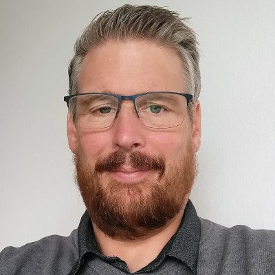
Dr. Lars Jansen
Sales Manager, Electron Microscopy


DAY 3: UHV- SCANNING PROBE MICROSCOPY

PROGRAM DAY 3

9:00 – 9:10Welcome & Agenda
9:10 – 9:30Introduction of Participants
9:30 – 9:55Martin Schaefer: Overview on UHV SPM Systems
9:55 – 10:30Steffen Porthun ( RHK Technology): The internals of the R10 SPM controlle
10:30 – 11:00Coffee Break
11:00 – 11:35Dr. Robert Ranecki, TU Kaiserslautern/ PAS Poznan/: 
SF-IETS investigations on metallacrown systems
11:35 – 12:10Dr. Rajan Adhikari, Univ. Erlangen/Schaefer Technologie GmbH:  
Scanning probe studies of porphyrins on metal surfaces
12:10 – 13:30Lunch Break
13:30 – 13:50Martin Schaefer: Cryogenic Nanopositioners
13:50 – 14:30Steffen Porthun (RHK Technology): Lock-Ins and PLLs
14:30 – 14:45Company Tour
14:30 – 16:30Coffee Break | Networking | Product Demonstrations

Host Day 3

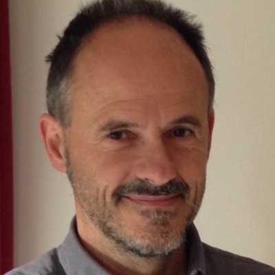
Dipl.-Phys. Martin Schaefer
Sales Manager


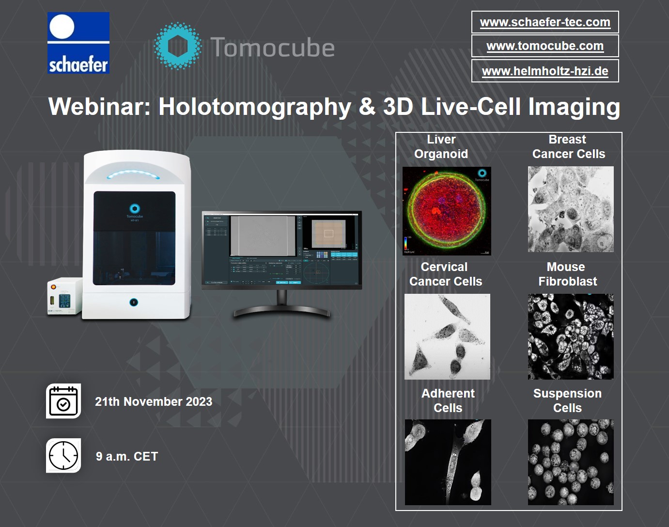
Webinar: Tomocube Label-Free 3D Holotomography & Imaging

Click Here to Register!

Visualization of living cells and organoids without labelling in high-resolution, in 3D and in real-time is now possible with Tomocube Holotomography solutions. Be with us in this Webinar to learn more about the revolutionizing potentials of this technology as a powerful tool for your research projects and applications.

Please don´t forget to register via the link. After entering your information, please click on “Send/Senden” botton below. The Webinar invitation link will be sent to you separatley after your registration.

👨‍🔬  Prof. Dr. Yongkeun (Paul) Park (KAIST & Tomocube CTO)

📅  Tuesday, 21 November 2023, 9 am CET


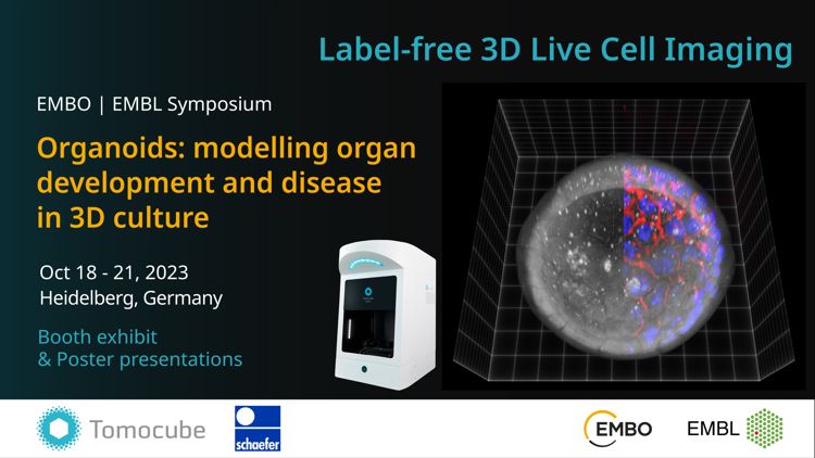
📢 Tomocube and Schaefer Technology at the EMBO | EMBL Symposium (II): Organoids!

Visualization of living organoids without labelling in high-resolution, in 3D and in real-time is now made possible with TOMOCUBE, INC. Holotomography -the HT-X1. Be with us at the EMBO | EMBL Symposium in Heidelberg to learn more about the revolutionizing potentials of organoids as a powerful research model, alongside the cutting-edge imaging technology that comes with it…

Registration for virtual attending is still available until Oct 11th. Don’t miss it. Click here for more information: Symposium II


📍 University of Heidelberg, Klaus Tschira Auditorium, Booth Org12

📅 18-21 October 2023


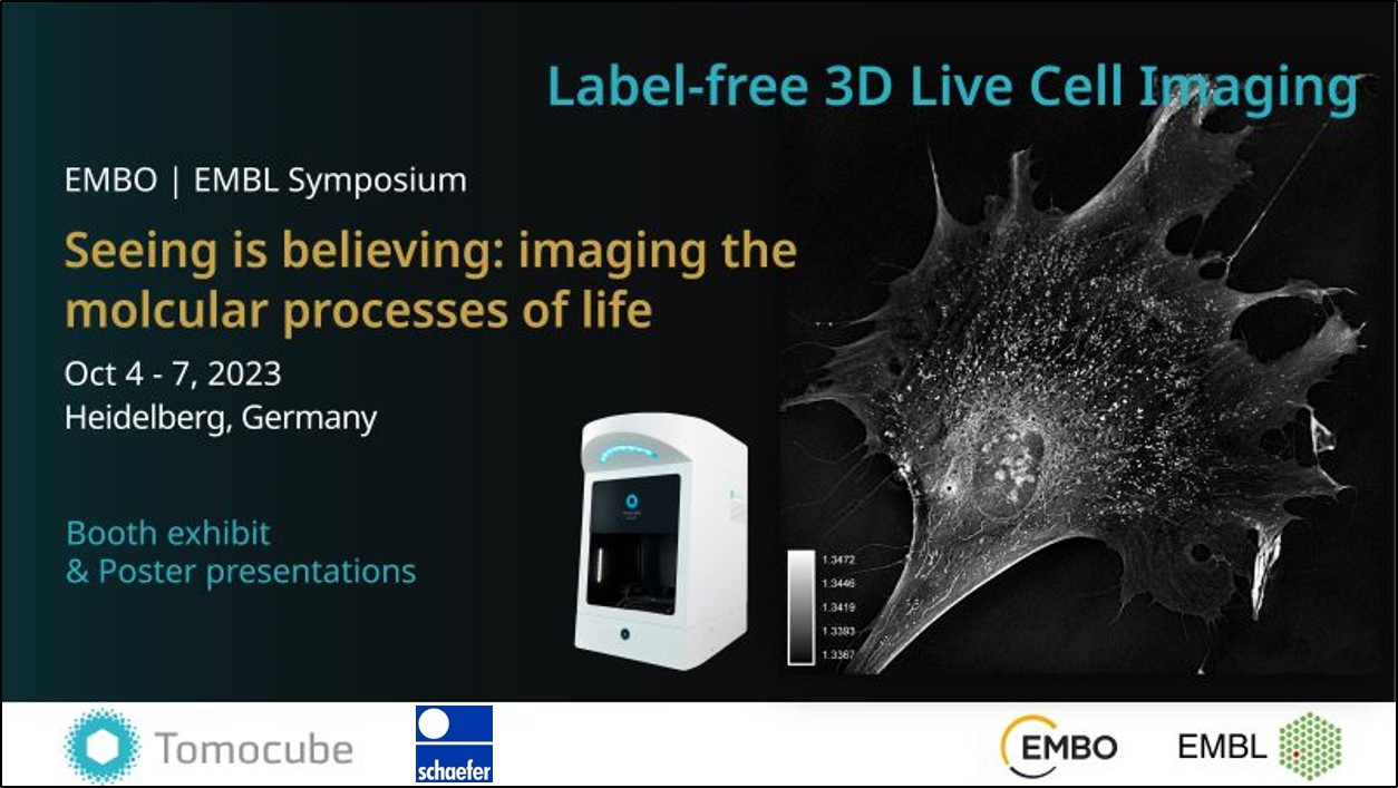
📢 Tomocube and Schaefer Technology at the EMBO | EMBL Symposium (I): Seeing is believing!

Visualization of living cells without labelling in high-resolution, in 3D and in time is no longer a challenge in cell biological research.

Be with us at the EMBO | EMBL Symposium in Heidelberg between Oct 4-7 to check out all the latest updates on imaging technology, biological research and data analysis. Because Seeing is Believing.

Registration for virtual attending is still available until Sep 27th. Don’t miss it. Click here for more information: Symposium I

📍 University of Heidelberg, Klaus Tschira Auditorium, Booth SiB05

📅 04-07 October 2023


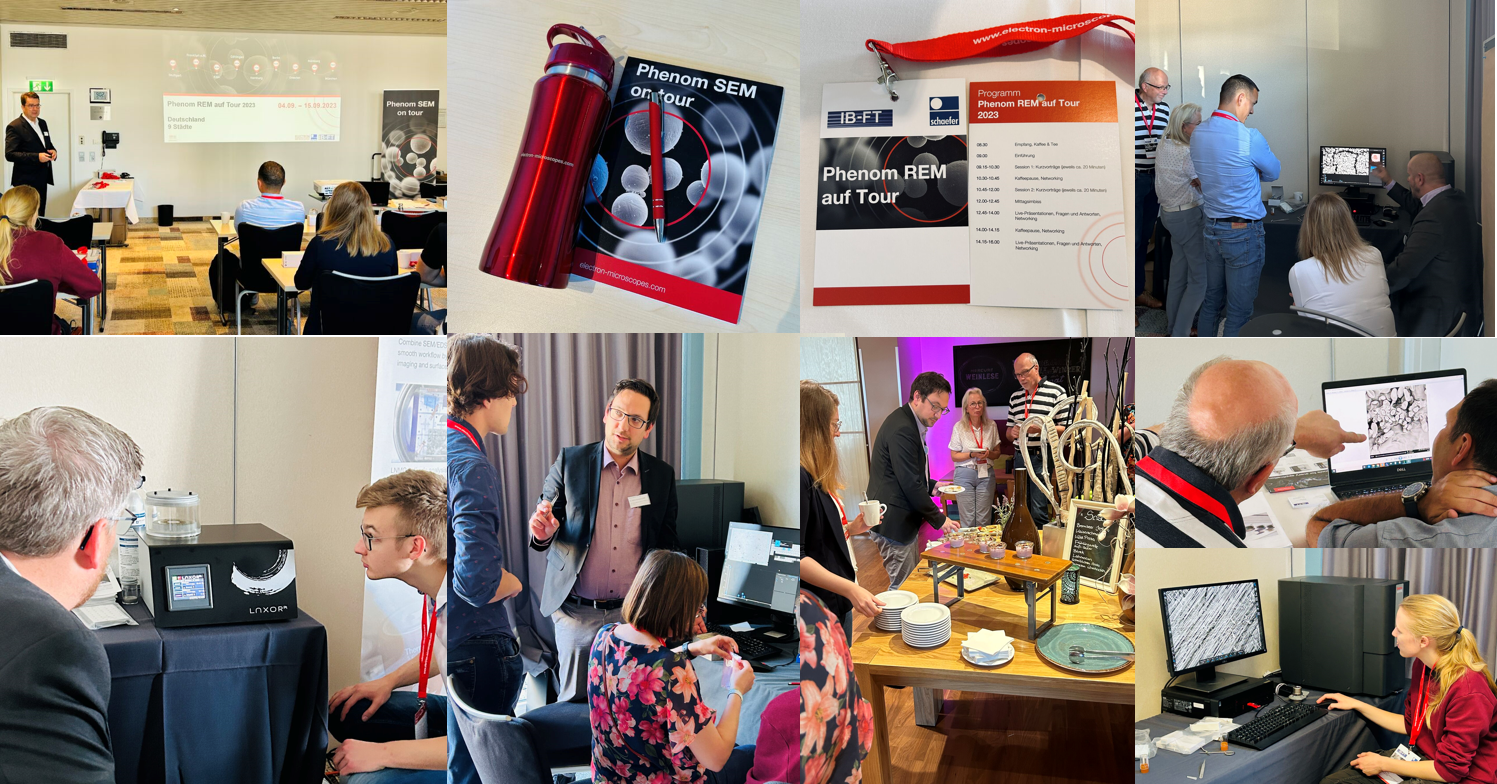
📢 Phenom SEM On Tour 2023: Our Successfull Roadshow Continues!

What an incredible display of energy, enthusiasm, and interest on the third day of the Phenom SEM Roadshow in Germany organized by IB-FT GmbH and Schaefer Technologie GmbH together with Thermo Fisher Scientific.

Next to the line-up of speakers from different fields, insightful presentations and live demonstrations on the ThermoScientific Phenom XL desktop SEM, Phenom ProX desktop SEM, LUXOR SEM COATING and Imina Technologies SA, attendees had the opportunity to witness real-time analyses of their own samples.

The feedback from participants thus far has been overwhelmingly positive, with many expressing their admiration for the wealth of insightful and relevant content presented. The event’s focus on delivering informative and valuable insights, rather than being overly commercial, has been particularly appreciated. Moreover, the certificate of participation provided at the end of each day is seen as a delightful addition and a testament to the seminar’s quality.

The Phenom SEM on tour continues its journey, with Göttingen as the next destination tomorrow and five more locations scheduled for the following week. After that, this electron microscopy event will extend its reach to several other countries, encompassing:

Belgium, by Benelux Scientific on 28.09 and 26.10.
Netherlands, by Benelux Scientific B.V. on 05.10 and 12.10.
France, by France Scientifique on 05.10, 26.10 and 16.11.
Austria, by Schaefer Technologie GmbH on: 17.10 and 19.10.
Switzerland, by Schaefer Technologie GmbH on 31.10 and 02.11.

So proud to see already results of this remarkable initiative by the companies within our group. Their collaborative efforts are making a substantial impact by providing valuable electron microscopy techniques to benefit a multitude of people.


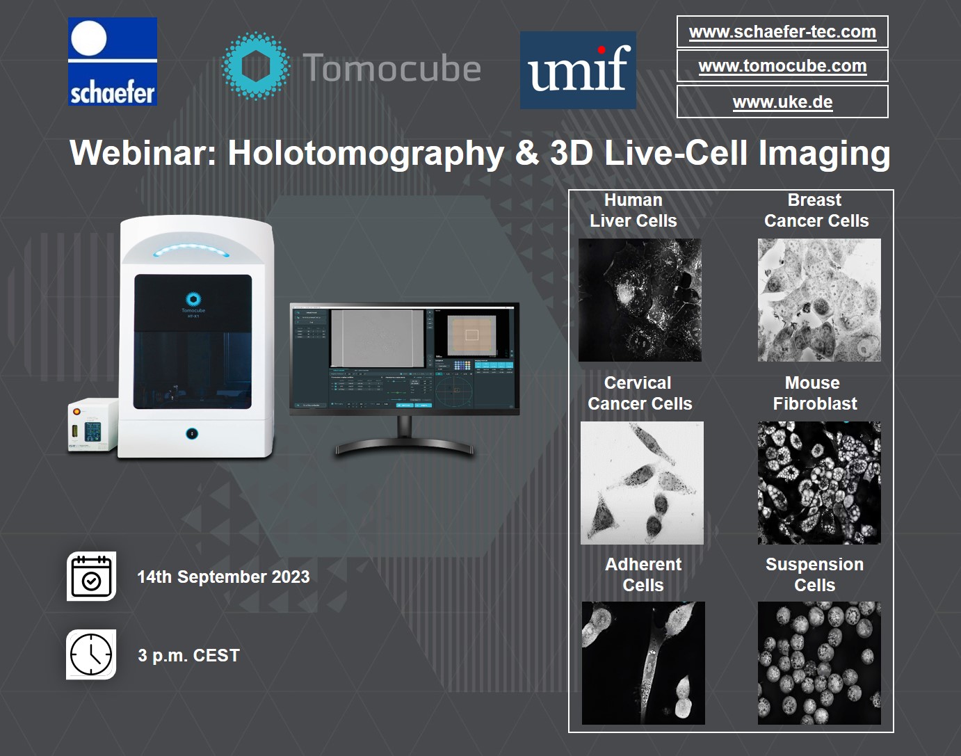
Webinar: Tomocube Holotomography & 3D Live-Cell Imaging!

Click Here to Register!


Holotomography (HT) enables imaging of cells and cellular organelles in 3D without using intrusive dyes or labelling. This novel technique prevents the perturbation of the cell physiology or morphology which in turn provides a true picture of the cell and its functionality. Label-free, live-cell imaging with quantitative measurement allows accurate 3D measurement of cell morphology, cellular interactions, and cellular kinetics.

We are proud to announce a new free joint free webinar in cooperation with umif in Hamburg presenting Tomocube holotomography solutions. Register and explore more about this unique technology in the field of life science research/applications.


📅 14th September 2023

⏲ 3 p.m. CEST


PHENOM SEM ON TOUR 2023

SEPTEMBER – NOVEMBER 2023

6 Countries, 20 Locations


Mark your calendar for your nearest, free, one-day seminar – focused on insights and hands-on experiences on innovative electron microscopy solutions. 

The biggest Phenom SEM on tour in Europe is set to start in September until November, covering 6 different countries with 20 different locations – bringing close to you the innovative technique of electron microscopy.   

These events are free of charge.

Click for further Information and Registration:

Germany  Austria  Switzerland (DE)  Switzerland (FR)  Belgium  Netherlands  France


Select your country

Wählen Sie Ihr Land | Sélectionnez votre pays

Select your country

Are you looking for products? Then select the country in which you are located or in which to deliver our products from the above list.

Wählen Sie Ihr Land

Sie suchen nach Produkten? Dann wählen Sie bitte aus der oben aufgeführten Liste das Land aus, in dem Sie sich befinden bzw. in das wir unsere Produkte liefern sollen.

Sélectionnez votre pays

Vous recherchez pour les produits? Alors s'il vous plaît sélectionner dans la liste ci-dessus? Mentionné le pays dans lequel vous vous trouvez ou dans laquelle nous voulons livrer nos produits.