Label-Free Quantitative Holotomographic Imaging: An Innovative Approach to Explore Cells and Tissues
Holotomography merges holography and tomography to solve the challenge of recovering light phase shifts to generate image contrast in Quantitative Phase Imaging, revealing intricate three-dimensional details within samples that would otherwise remain hidden with traditional techniques. Just as a CT scan uses X-ray absorptivity as the imaging contrast to see inside a patient’s organs without invasive procedures, HT uses the refractive index (RI), an intrinsic optical parameter describing the speed of light passing through a specific material, to visualize living cells and tissues. Since variations in the biomolecular concentration directly impact the overall RI of the cellular biomaterials, the reconstruction of RI tomogram by Holotomography can retrieve important biophysical parameters such as cell volume, surface area, or protein concentration for further analysis. In general, all Holotomography systems consist of three essential components: (1) illumination optics to capture structural information of the specimen in light wave; (2) imaging optics to encode information in light onto a 2D intensity image; and (3) a reconstruction algorithm to decode and synthesize the raw data into a 3D tomogram. Holotomography’s advantages lie in its capability to provide non-invasive, label-free 3D images of live cells, allowing real-time studies while the measurement of RI tomogram also allows quantitative analysis. Its data acquisition speed surpasses traditional methods, making it invaluable for quick results in medical diagnostics and drug research. Holotomography’s applications span from cellular components like nuclei and mitochondria to studying organoids, tissues, and even whole organisms, enabling breakthroughs in cellular biology understanding. Its applications continue to expand as new advances are made in tomographic imaging.
Tomocube Holotomographic Microscopes
HT-2H
HT-X1
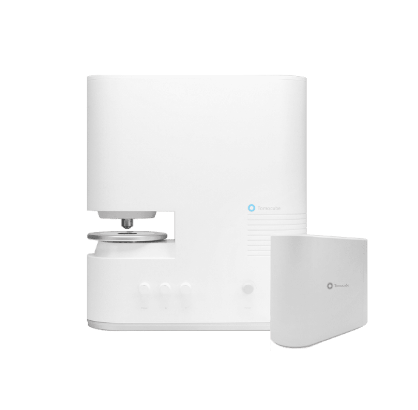
HT-X1™
The #1 Choice for Live-Cell Imaging and Analysis Empowering Your Exploration. Redefining the limits.
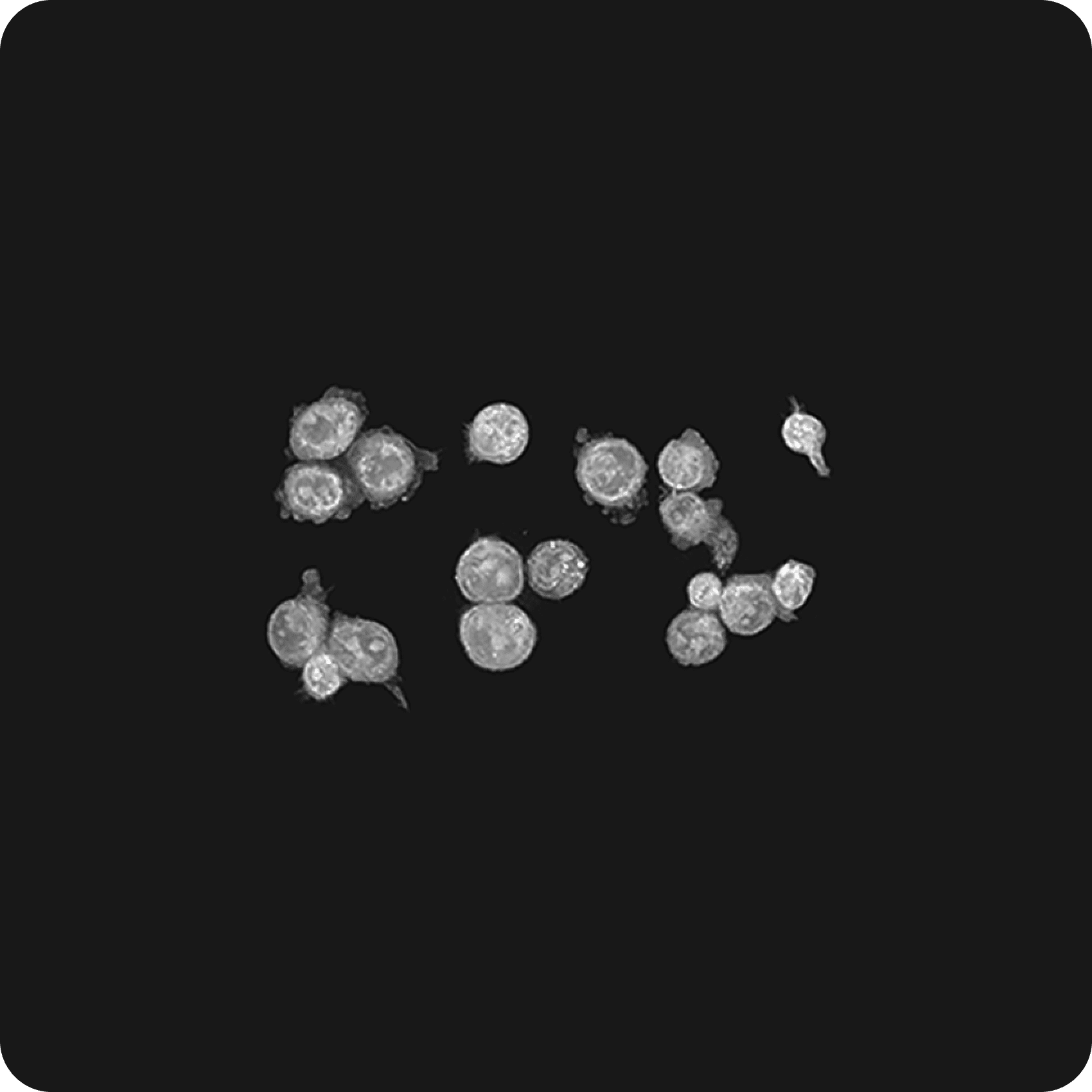
Holotomography is a technique that utilizes low-intensity light to acquire the refractive index of cells from multiple angles. This technology has emerged as an excellent cellular imaging solution, allowing for high-resolution imaging while maintaining the health of the cells by using low-intensity light sources. Tomocube’s second-generation Holotomography, the Tomocube HT-X1, employs low-coherence light sources, ensuring lower toxicity and noise-free high-resolution image acquisition compared to laser-based methods.
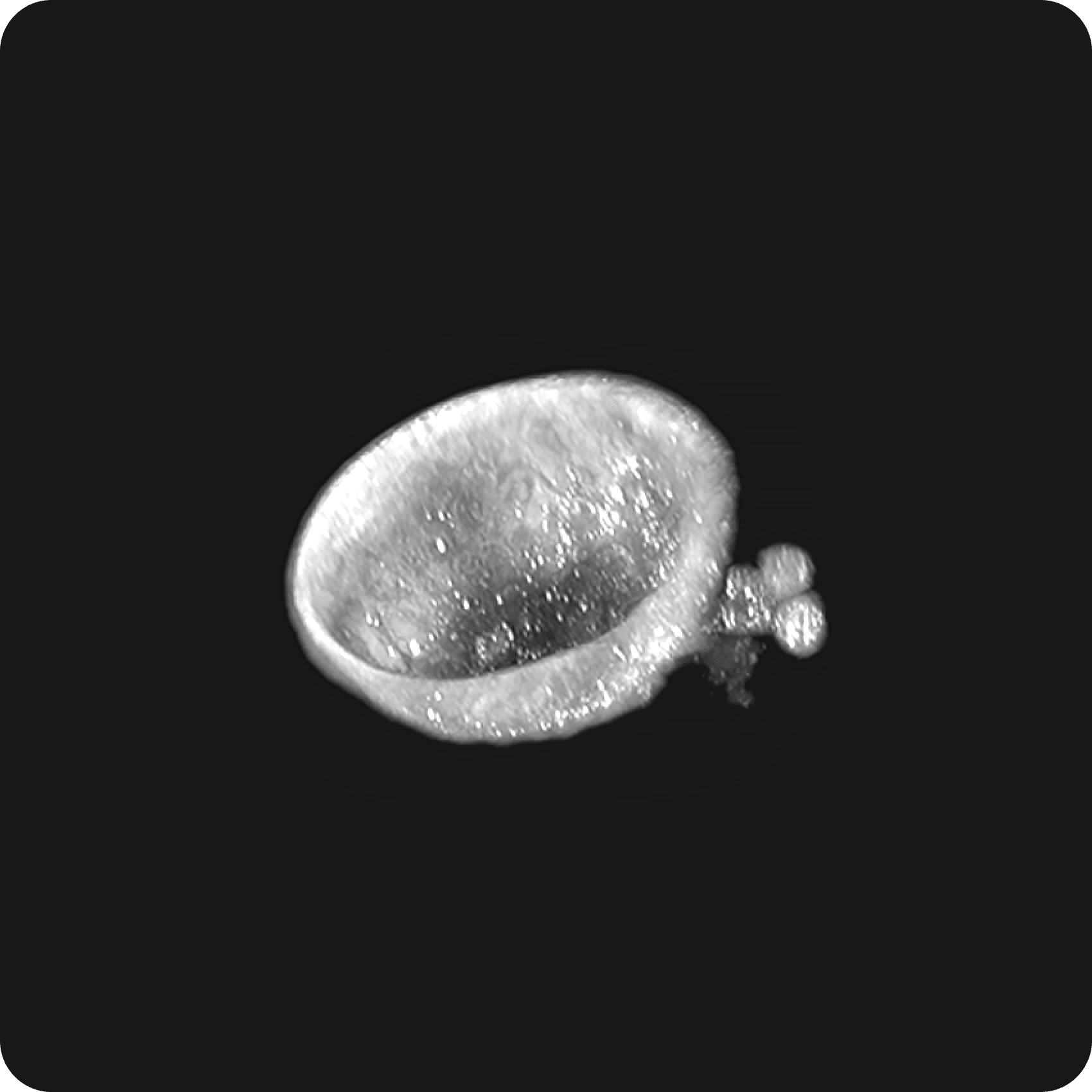
The high-resolution images obtained through this technology enable real-time observation of not only the morphology of living cells but also the shapes of subcellular organelles, such as the nucleus, nucleolus, mitochondria, and lipid droplets. The integrated stage-top incubator provides a stable cultivation environment, enabling prolonged monitoring of sensitive cells like stem cells or organoids.
This innovative system is compatible with various commercial imaging plates and supports imaging up to 96-well plates, making it applicable for high-throughput screening. Utilizing the HT-X1 allows for the acquisition of high-resolution images of cells, microorganisms, organoids, and tissue samples. Additionally, Holotomography analysis software, TomoAnalysis, facilitates obtaining quantitative information based on refractive index, including high-resolution 3D images of cells, as well as volume, area, concentration, and dry mass. Therefore, the use of HT-X1 becomes a crucial tool in understanding biological phenomena.
Product Features
High resolution, high contrast and high sensitivity
Large-field detection
Programmable multi-well analysis
Laser autofocus for plate-to-plate reproducibility
Low-noise, stain-free imaging without further calibration steps
Quantitative Bioimaging
Integrated incubation for long-term time-lapse
Integrated 5-channel FL light engine
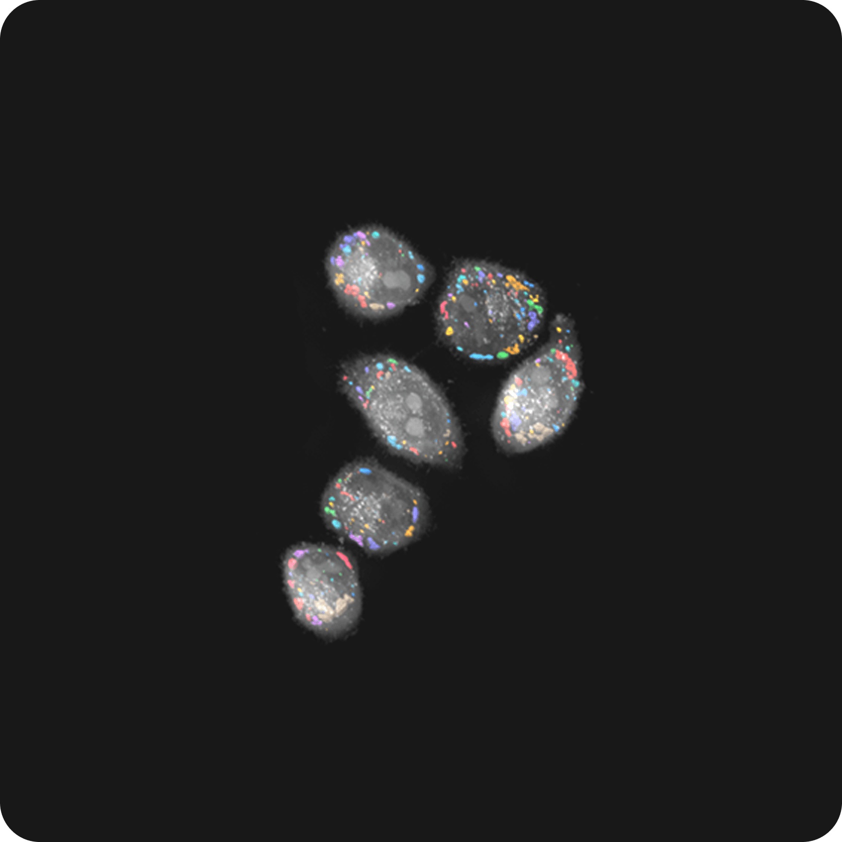
Correlative holotomography with 3D fluorescence
True multidimensional imaging across time, space and modality
HT-X1 Introduction
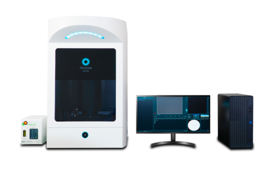
HT-2H
Revolutionary Holotomography opens a new era in label-free 3D live cell imaging
the field of advanced imaging technology, Tomocube’s very first generation of Holotomography stands out as a symbol of innovation. Leveraging groundbreaking 3D quantitative phase imaging technology, the HT-2H employs diffraction tomography to produce high-resolution 3D holographic images of unlabeled live cells and tissues. This innovative imaging series enables the rapid and straightforward acquisition of real-time, nanoscale images of individual cells, without the need for sample preparation.
By reconstructing a 3D distribution map of the refractive index of biological specimen from multiple 2D holograms, HT-2H provides researchers with essential insights into various cellular and subcellular organelle characteristics, such as volume, shape, cytoplasmic density, surface area, or deformability. Meanwhile, its long-term time-lapse imaging capability enables researchers to observe dynamic changes within samples over extended periods.
Additionally, the HT-2H seamlessly integrates the two complementary modalities of Holotomography and fluorescence imaging, excelling in its ability to offer long-term tracking of specific targets while minimizing unwanted stress to cells. Winner of the Microscopy Today 2019 Innovation Award, the HT-2H facilitates broader access for researchers and clinicians to quantitatively investigate cell pathophysiology and assess the effectiveness of drug interventions.
Product Features
Fast and label-free imaging
High-resolution (super-resolution) without moving parts
Long-term live-cell imaging
Negligible photo damage due to low power laser
Real-time analysis with quantitative data
Upgradeable with 3D fluorescence imaging capability
Holotomography with Tomocube
TomoAnalysis™
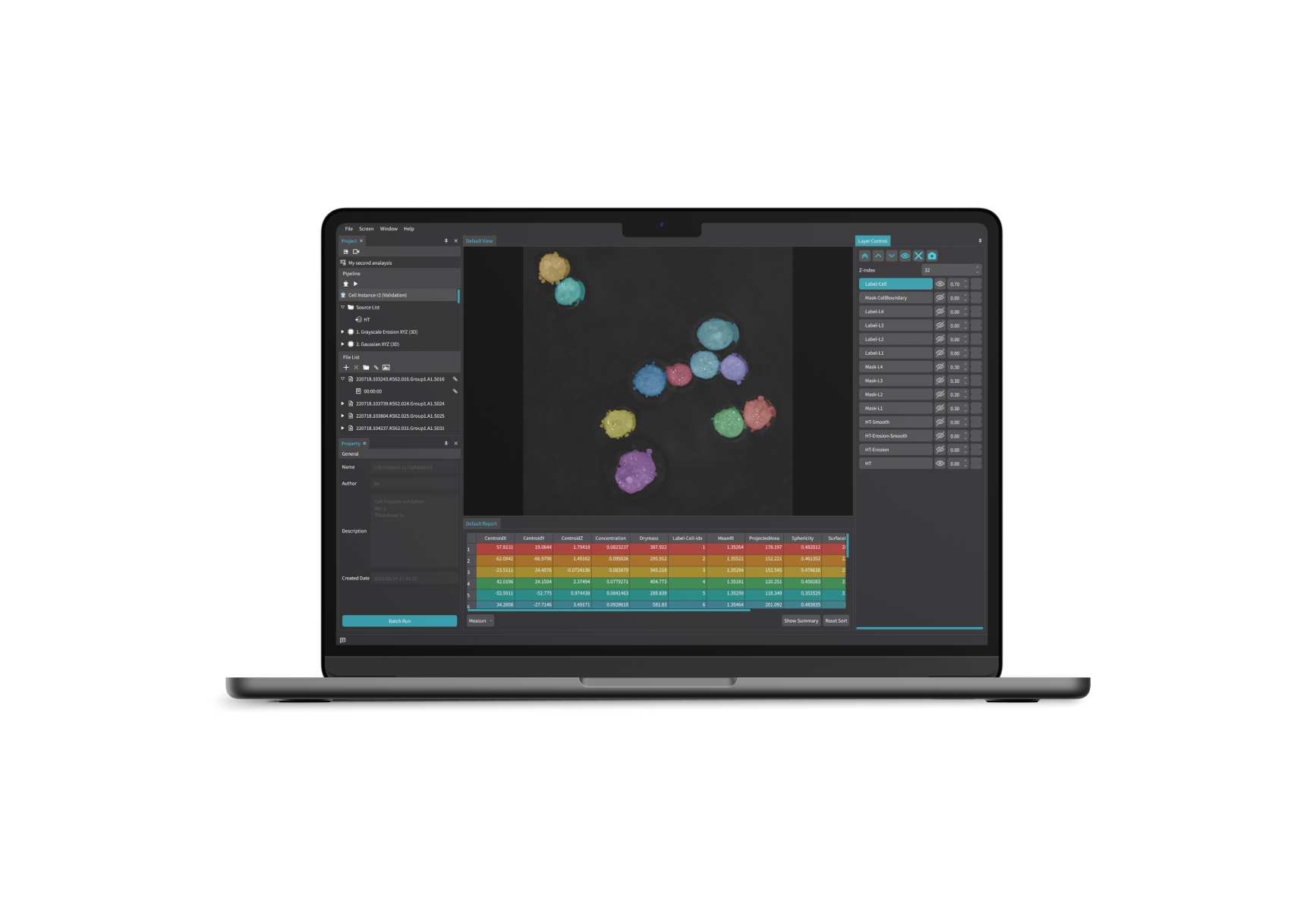
Unleash the Full Potential of Holotomography
Tomocube’s TomoAnalysis offers advanced capabilities for the visualization and analysis of refractive index(RI) tomograms down to the cellular and subcellular levels. Through a combination of different segmentation strategies and AI-driven deep learning models, the software provides precise quantification results of important parameters such as volume, surface area, sphericity, aspect ratio, and RI concentration of the regions of interest, facilitating a comprehensive understanding of the specimen properties.
With a range of customizable analysis pipeline procedures designed to address diverse user needs, researchers can refine their multimodal image data, extract meaningful measurements, and conduct in-depth quantitative analyses.
Are you looking for products? Then select the country in which you are located or in which to deliver our products from the above list.
Wählen Sie Ihr Land
Sie suchen nach Produkten? Dann wählen Sie bitte aus der oben aufgeführten Liste das Land aus, in dem Sie sich befinden bzw. in das wir unsere Produkte liefern sollen.
Sélectionnez votre pays
Vous recherchez pour les produits? Alors s'il vous plaît sélectionner dans la liste ci-dessus? Mentionné le pays dans lequel vous vous trouvez ou dans laquelle nous voulons livrer nos produits.