Webinar: Novel AI-Assisted Tissue Image Analysis Software from TissueGnostics

Register Now!


We are proud to announce a new webinar presenting TissueGnostics‘s analysis capabilities. We will walk you through all capabilities.

đź“… 16th June 2023
⏲ 10:00 a.m. Amsterdam, Berlin, Rom, Stockholm, Vienna

What You will Learn:

1. Context-based analysis 

  • Analyze tissue sections, TMAs, confluent cells, smears
  • Nuclear segmentation, total area measurements, dot detection
  • Cytoplasmic and membrane measurements

2. Streamlined workflow 

You’re only 4 simple steps away to your final results:

  • Automated segmentation
  • Quantitative analysis
  • Data verification
  • Results exportation

3. Also available as a standalone software

Wether or not you are needing a new microscope TissueGnostics has the solution for you. From slide loaders to confocal microscope we have it all!


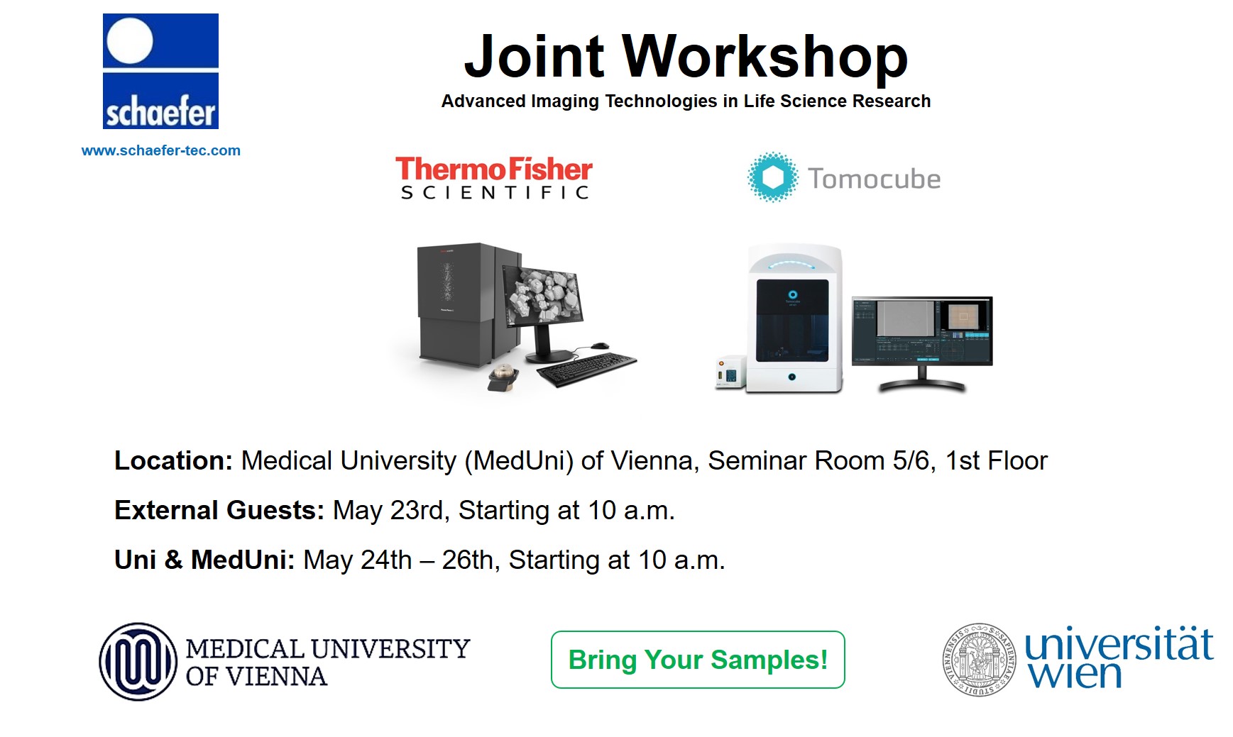
Workshop: Advanced Imaging Technologies in Life Science Research

What You will Learn:

ThermoFischer Desktop STEM: Scanning transmission electron microscopy (STEM) systems have for some time been available as an additional modality for TEM systems, promising increased contrast, particularly from unstained sections. But they have not been widely available outside of specialist comprehensive EM centers. In this webinar, we discuss the advantages of low-kV STEM imaging using the highly automated Thermo Scientific™ Phenom Pharos™ G2 FEG-SEM with the new scanning transmission electron microscopy (STEM) detector, which can achieve a higher resolution than the BSD detector at only 15 kV.

Tomocube Holotomography: Holotomography (HT) allows for the imaging of cells and cellular organelles in 3D without the use of intrusive dyes or labelling. This novel technique prevents the perturbation of the cell physiology or morphology which in turn provides a true picture of the cell and its functionality. Label free, live cell imaging with quantitative measurement allows accurate 3D measurement of cell morphology, cellular interactions, and cellular kinetics. This webinar will discuss the technical aspects of how holotomography works, its advantages and its limitations. We will illustrate showing examples of what is possible.


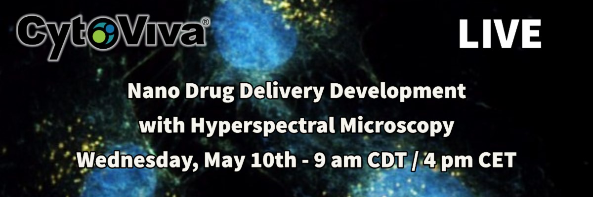
Webinar: Nano Drug Delivery Development with Hyperspectral Microscopy

Register Now!


What You will Learn:

CytoViva’s Hyperspectral Microscopy is a highly effective tool for the development of nanoscale vectors for drug delivery. It not only provides label free imaging of these nanoscale vectors and their active pharmaceutical ingredients (API), but also enables spectral confirmation of these materials in cells and tissue, including spheroids and organoids.

In this webinar you will learn the principles behind Enhanced Darkfield Hyperspectral Microscopy. This will include an overview of nanoscale darkfield microscopy and high-resolution hyperspectral imaging, which produces optical spectrum in each nanoscale pixel of an image.

Additionally, various nano-vector drug delivery applications will be reviewed. This will include data illustrating the confirmation of APIs on individual nanoparticles along with label free imaging and spectral confirmation of nano-vectors and their drug load in cells and tissue. Examples showing API distribution in larger scale samples will also be demonstrated. Join us to learn how you can use darkfield hyperspectral microscopy to advance your work developing nano drug delivery vectors.


Byron Cheatham, Vice President @ CytoViva, Inc., USA

Since 2005, CytoViva has helped hundreds of research and industry laboratories solve critical problems at the nanoscale with its state of the art hyperspectral microscope technology. Research applications supported by CytoViva range from nanotechnology to digital pathology.


Webinars: Advanced Imaging Technologies in Life Science Research

Date: May 11th, 2023

ThermoFisher: 2 – 2:30 pm CEST

Tomocube: 2:30 – 3:30 pm CEST

Event Link: Click Here to Participate


What You will Learn:

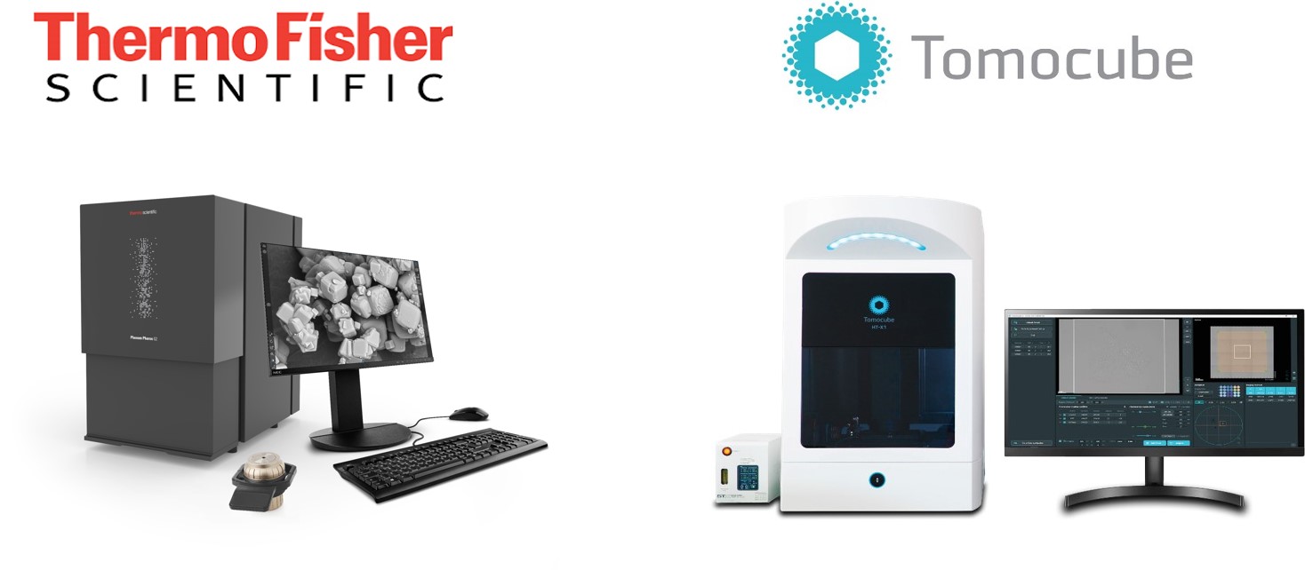
ThermoFischer Desktop STEM (Webinar 1): Scanning transmission electron microscopy (STEM) systems have for some time been available as an additional modality for TEM systems, promising increased contrast, particularly from unstained sections. But they have not been widely available outside of specialist comprehensive EM centers. In this webinar, we discuss the advantages of low-kV STEM imaging using the highly automated Thermo Scientific™ Phenom Pharos™ G2 FEG-SEM with the new scanning transmission electron microscopy (STEM) detector, which can achieve a higher resolution than the BSD detector at only 15 kV.

Tomocube Holotomography (Webinar 2): Holotomography (HT) allows for the imaging of cells and cellular organelles in 3D without the use of intrusive dyes or labelling. This novel technique prevents the perturbation of the cell physiology or morphology which in turn provides a true picture of the cell and its functionality. Label free, live cell imaging with quantitative measurement allows accurate 3D measurement of cell morphology, cellular interactions, and cellular kinetics. This webinar will discuss the technical aspects of how holotomography works, its advantages and its limitations. We will illustrate showing examples of what is possible.


Select your country

Wählen Sie Ihr Land | Sélectionnez votre pays

Select your country

Are you looking for products? Then select the country in which you are located or in which to deliver our products from the above list.

Wählen Sie Ihr Land

Sie suchen nach Produkten? Dann wählen Sie bitte aus der oben aufgeführten Liste das Land aus, in dem Sie sich befinden bzw. in das wir unsere Produkte liefern sollen.

SĂ©lectionnez votre pays

Vous recherchez pour les produits? Alors s'il vous plaît sélectionner dans la liste ci-dessus? Mentionné le pays dans lequel vous vous trouvez ou dans laquelle nous voulons livrer nos produits.