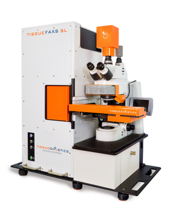
Automated Cutting-Edge Tissue Cytometry for Future-Oriented Quantitative Biomedicine
The high-end flexible tissue cytometer “TissueFAXS” provides high-precision biomedical microscopic solutions with AI-assisted image analysis tools (e.g. Machine/Deep Learning) enabling comprehensive phenotypical and functional analyses (e.g. FACS) in tissue context and on the single cell level. The classification and nuclei segmentations even in dense tissues are no longer a considerable challenge. Image acquisitions include brightfield, fluorescence, confocal and multispectral analyses. The high-throughput automated loading and whole-slide scanner facilitates up to 120 standard slides or 60 double-sized slides. The inverted system allows live-cell and time-lapse imaging of samples in petri dishes, well plates and cell culture flasks. The TissueFAXS platform is available in various hardware and software configurations suitable for different research activities.
Product Features
- High-resolution imaging
- High-content phenotyping
- Multispectral imaging and spectral unmixing
- Imaging mass cytometry
- Spatial relationships
- Live-cell imaging
- Single-cell detection and contextual image analysis
- High-dimensional co-expression analysis
- FISH, seqFISH, and CISH detection
Introduction of TissueGnostics Introduction TissueFAXS Q Platform
Click and Learn about Different TissueFAXS Configurations
Brightfield Cytometer


Fluorescence Cytometer


Brightfield + Fluorescence Cytometer


Confocal Cytometer Multispectral Cytometer


Click and Learn about Different TissueFAXS Software Solutions
Scanning & Viewing


Single Cell Analysis
HistoQuest
Contextual Image Analysis
Applications in Human, Animal and Plant Tissues
- Immunophenotyping of organs/tissues in-situ
- Spatial phenotypic and functional characterization of tumor- and cellular-microenvironment
- Identification of single cells even in dense tissue
- Automated classification of tissue compartments
- Multiplexing and spectral unmixing to perform high-plex assays on histological sections with single-cell precision
- Structural analysis and determination of subcellular marker localization
- Molecular single-cell profiling using antibody labeling, FISH, CISH, RNA-ISH, and RNAScope
- Quantification of cellular pathogens and intracellular parasites
- Panoramic imaging and quantitative histopathology of whole-slide biopsies, TMAs and even big tissue sections


