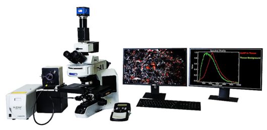
CytoViva® Darkfield- and Hyperspectral Microscopy
CytoViva’s enhanced darkfield optics allows the visualization of nanoscale objects (metals, oxides, polymers, viruses or liposomes) without any fluorescent labeling. A spectrograph (VNIR 400-1000nm oder SWIR 900-1700nm) acquires hyperspectral images from which the nano-materials can be characterized and classified.
The CytoViva® technology works in different substrates and environments (e.g. cells, tissue, all kinds of liquids). The hyperspectral analysis is very sensitive on chemical properties of the refracting objects and can for example resolve different surface modifications (functional groups) individually on single nanoparticles.
CytoViva® Measurement and Analysis
- High-resolution darkfield image with very high signal to noise ratio
- Line-by-line hyperspectral image acquisition with X-Y scan stage and spectrograph
- Generation of spectral libraries of the targeted specimen
- Spectral angular mapper procedure to map the matching data in the hyperspectral image
CytoViva® Introduction CytoViva® Webinar
Click and Learn more about Different CytoViva® Products
CytoViva® Enhanced Darkfield Microscopy


CytoViva® Hyperspectral Microscope

CytoViva® 3D Enhanced Darkfield Imaging

CytoViva® Integrated Raman & Hyperspectral Microscopy

Click and Learn more about Different CytoViva® Applications


Nanoparticle Darkfield Hyperspectral Imaging Digital Pathology Nano-Drug-Delivery

Microbiology & Virology
Further Applications:
- Nanotoxicity of particles in cell cultures
- Determination of functional groups of Drug Delivery particles
- Drug Loaded Liposomes
- Particles for biosensors
- Microplastic in the environment and in tissue
- Implant debris in tissue
- Carbon Nanotubes
- Wafer defect analysis


Further Links:
- Diverse application examples on the CytoViva Youtube channel
- Published results to compare to REM-EDS and Raman-spectroscopy www.ncbi.nlm.nih.gov/pubmed/26864497 and as video http://www.jove.com/video/53317
System Options:
- Dual Mode Fluorescence for simultaneous observation of fluorescent and scattering sample areas
- 3D Enhanced Darkfield Imaging
- Makro-system for wafer defect analysis